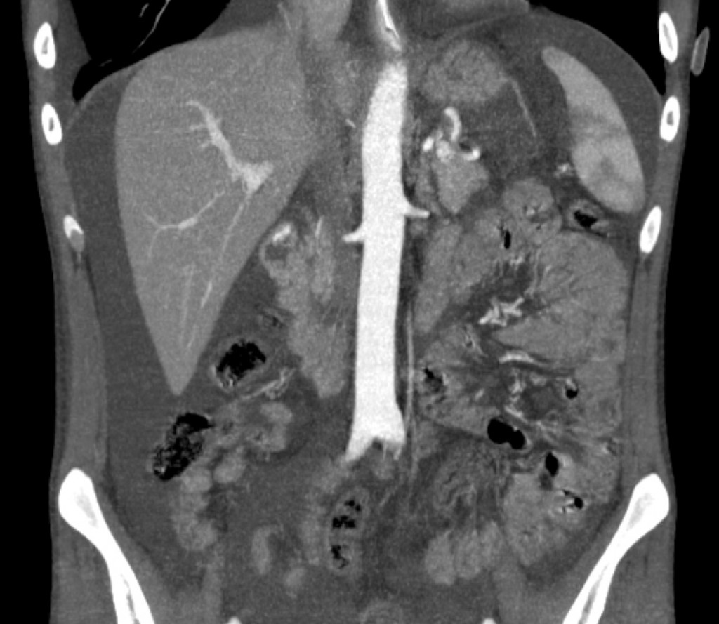

Острое дуоденальное кровотечение
Анамнез: Мужчина, 24 года. Страдает ХПН. Острая анемия тяжелой степени, источник кровопотери не известен. При фиброгастроскопии (осмотр до начальных отделов луковицы 12-перстной кишки) и фиброколоноскопии источника кровотечения не найдено.

Обсуждение/выводы:
"CT angiography typically detects active bleeding with a rate that exceeds 0.3–0.5 mL/min, a rate that is slightly higher than the threshold assigned to catheter angiography (0.5 mL/min) and is lower than that of scintigraphy with 99mTc-labeled red blood cells, which can be used to detect active bleeding at a rate higher than 0.1 mL/min. Therefore, severe bleeding episodes, such as those manifesting with hemodynamic instability, increase the pretest probability of a positive result for active bleeding at CT angiography".
"Although the specific technical parameters used and the phase images acquired vary among institutions, most agree that three-phase examinations that include the unenhanced, arterial, and portal venous phases provide the best and most reproducible results for this clinical application in patients with acute gastrointestinal bleeding".
Литература
- Artigas JM, Martí M, Soto JA, Esteban H, Pinilla I, Guillén E. Multidetector CT angiography for acute gastrointestinal bleeding: technique and findings. Radiographics : a review publication of the Radiological Society of North America, Inc. 33 (5): 1453-70. doi:10.1148/rg.335125072 - Pubmed